
Advanced Technologies
At Emerald Eyes we offer only the best when it comes to our patients' care. We are equipped with the most advanced, state-of-the-art technology there is when it comes to assessing your vision and eye health. Our access to these advanced technologies is one of the many things that sets us apart from other eye doctors in the area.
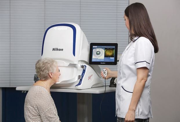
OPTOMAP®
Optos’ core product is the retinal image produced by the Panoramic200 Scanning Laser Ophthalmoscope device: the optomap® retinal image.
The optomap® retinal image gives eye-care professionals a much larger view (200 degrees) of the back of the eye – your retina – than conventional eye exam equipment. The images can be taken without dilating your pupils – a very common procedure which is uncomfortable and inconvenient for many people.
The optomap® image is captured in less than a second and is immediately available for doctor and patient to review. The optomap® Retinal Exam offers many clinical, practice and patient benefits.
The optomap® image is displayed immediately after being taken, allowing the eye care professional to review it quickly and if necessary, refer you to a retinal specialist. Using the internet, the image can be sent anywhere in the world for a specialist to review.
Each optomap® image is as individual as fingerprints or DNA and can provide eye care professionals with a unique view of your health very quickly and comfortably. The optomap® image is captured in less than one second and is immediately available for you and your doctor to review.
The optomap® retinal image offers many advantages including:
- An ultra-widefield view of the retina
- Comfortable and quick image capture
- Non-invasive. Eliminates need for dilating drops
- Helps you understand your eye health
- Provides permanent records for future comparison
- The Optomap® technology does not require pupil dilation, however the decision to dilate or not is a medical decision to be made by your health care professional
- Patient can resume normal activities immediately


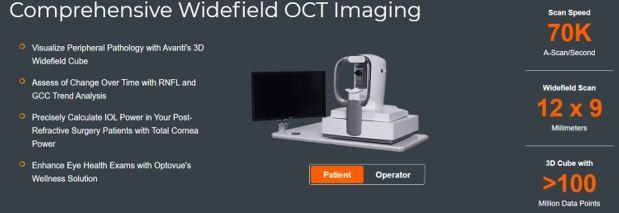
Optovue
- Visualize Peripheral Pathology with Avante's 3D Widefield Cube
- Access of Change Over Time with RNFL and GCC Trend Analysis
- Precisely Calculate IOL Power in Your Post-Refractive Surgery Patients With Total Cornea Power
- Enhance Eye Health Exams with Optovue's Wellness Solution
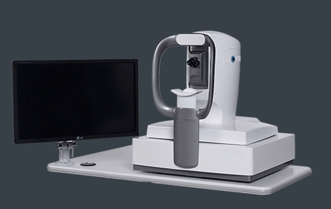
Oculus K5
A New Way to Treat Dry Eye Disease
The OCULUS Keratograph® 5 is a groundbreaking piece of imaging equipment that changes the way we diagnose and treat dry eye disease. This brand new machine is among the latest advances in optometry, it is especially useful for testing, diagnosing, and treating dry eye disease. It offers the following benefits over more complicated alternatives:
- Non-invasive way to take high resolution images
- Offers a wide variety of different tests and software analysis
- Gives patients more insight into their conditions and needs
What is the OCULUS Keratograph® 5?
The OCULUS Keratograph® 5M integrates a high definition camera, keratometer, and corneal topographer into one seamless and state-of-the-art imaging machine. Surface eye diseases are no match for this precise and innovative tool, which integrates with computer technology to create detailed, full-color digital images of your eye.
If you have dry eye disease, we will use the round camera lens to examine your Meibomian glands (also called your tarsal glands). These glands produce the oil layer of your tear film, which keeps them from drying out and evaporating. The Keratograph allows us to test the break-up time of your tear film, the height of your tear meniscus, and the condition of your eye’s lipid layer.
What You Should Know
Light-emitting diodes make it possible to create these highly complex images, while thousands of measuring points make our diagnoses more accurate and precise than ever. The Keratograph even relies on an infrared ring to prevent the involuntary secretions associated with bright lights and glares. Together, these tools work to create precise and useful records of the exact shape, size, function, and abnormalities of your eyes.

lipiflow
Designed To Improve Gland Function
The LipiFlow Thermal Pulsation System, a cleared medical device for Meibomian Gland Dysfunction (MGD), consists of a Console and a single-use sterile device, known as the Activator, and has a drug-free mechanism of action. Eye care professionals use the LipiFlow System to treat MGD patients in-office with confidence and efficiency.
The LipiFlow System represents more than 10 years of dedicated research and is protected by more than 30 patents. a phased pressure profile with adaptive force equalization and proximal-to-distal peristaltic motion evacuates gland contents as the inner lid is gently heated.
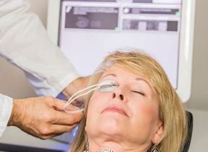
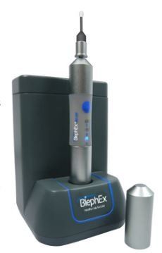
blephex
How BlephEx Works
BlephEx is a painless in-office procedure performed by your eyecare professional or trained technician. A revolutionary new patented BlephEx handpiece is used to very precisely spin a soft medical grade micro-sponge along the edge of your eyelids and lashes. This procedure effectively removes years of accumulated bacterial biofilm, much like a dental cleaning removes accumulated plaque, also a biofilm. Once the biofilm is removed, your eyelids are cleaner and healthier. A healthy eyelid is critical for normal tear production.
The patented micro-sponge is disposable and for optimal cleaning and exfoliating, a fresh clean one is used for each eye. The eyes are rinsed well afterwards. The procedure lasts about 6-8 minutes. Most patients simply report a tickling sensation. A numbing drop is usually placed in each eye prior to treatment for increased comfort.
After the procedure, you may resume normal activities. Home cleaning regimes can continue but are only semi-effective. BlephEx should be repeated every 4-6 months as determined by your eye doctor. The sooner in life BlephEx is begun as a routine element of eye care, the better the long-term results. If you wait until you have symptoms before seeking treatment, much damage may already be done yo your tear glands. Treat your eyelids early, and treat often.
prokera
Amniotic membrane is part of the placenta and is the tissue closest to the baby throughout development in the womb. Amniotic membrane protects the baby from any harm and has natural biologic actions which help the baby develop. The tissue has biological properties that aid in ocular surface repair.
The amniotic membrane tissue in PROKERA has natural therapeutic actions that help damaged eye surfaces heal. Eyes treated with PROKERA have less scarring and less inflammation. The amniotic membrane in PROKERA is thin and clear like the tissue on the surface of your eye and protects your eye’s damaged tissue while inserted.